Skip to content
The Initial Challenge: A Matrix of Anomalies
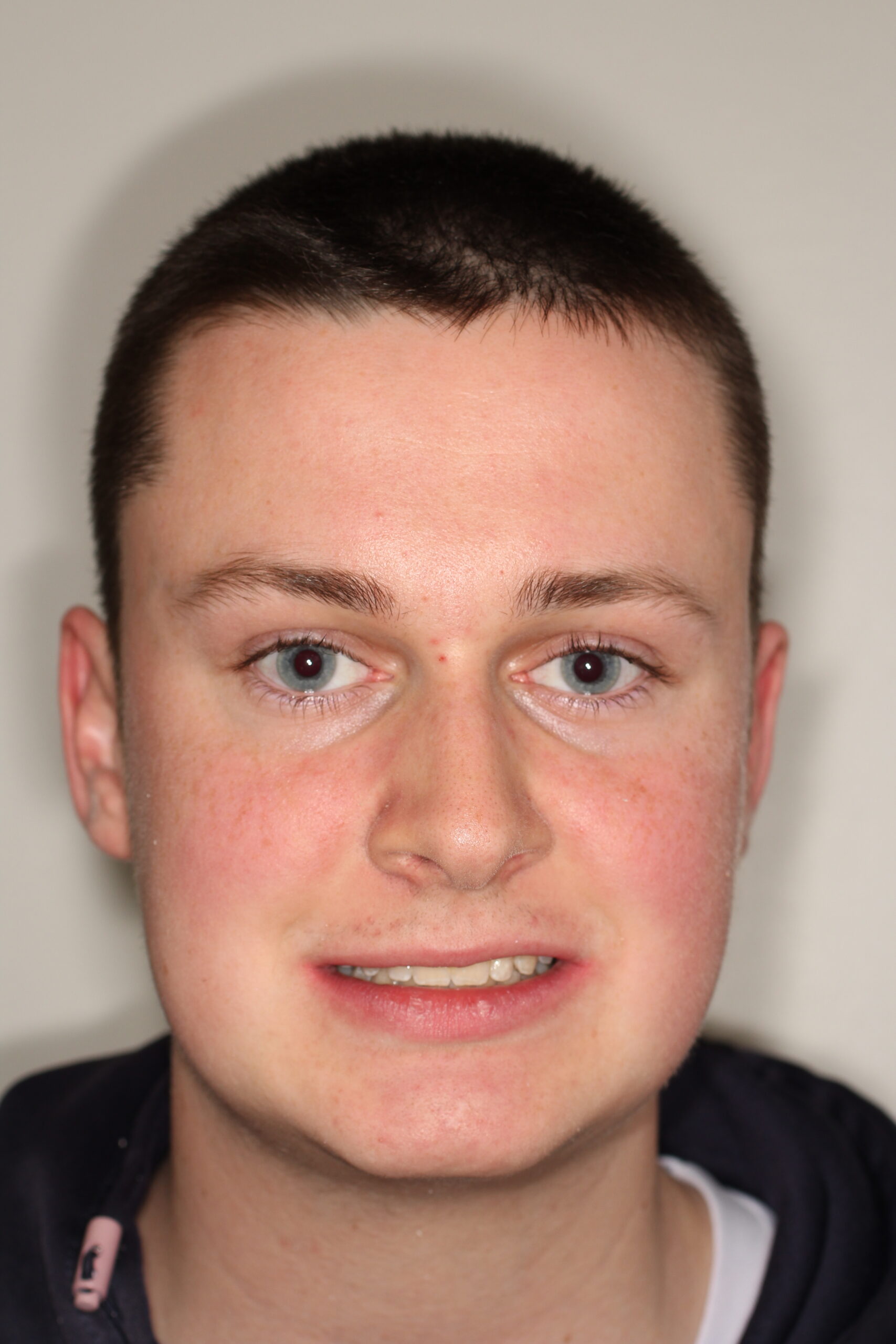
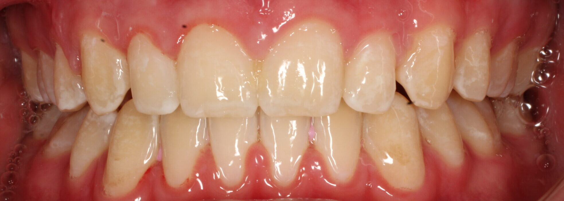
Matt’s initial clinical presentation involved a complex combination of structural and developmental hurdles that required deep investigation:
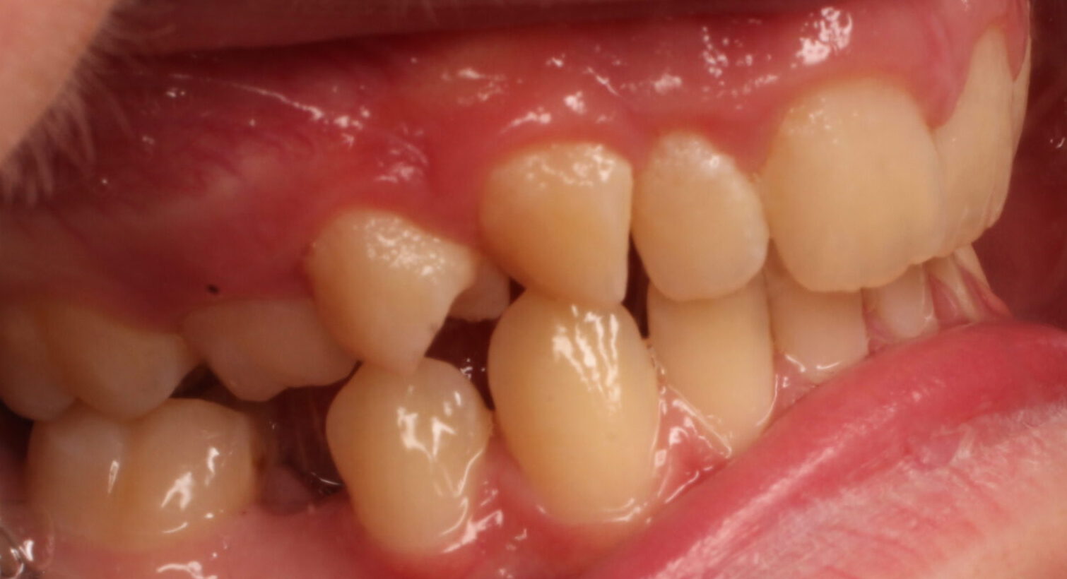
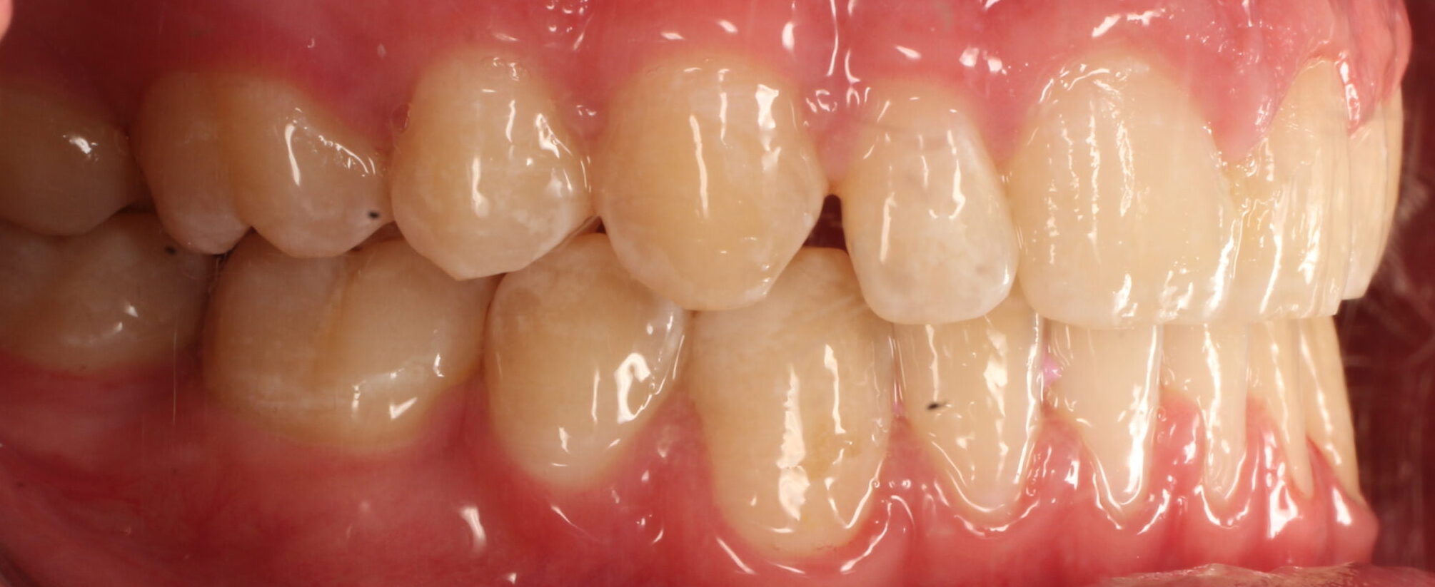
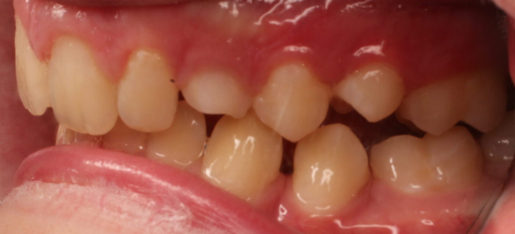
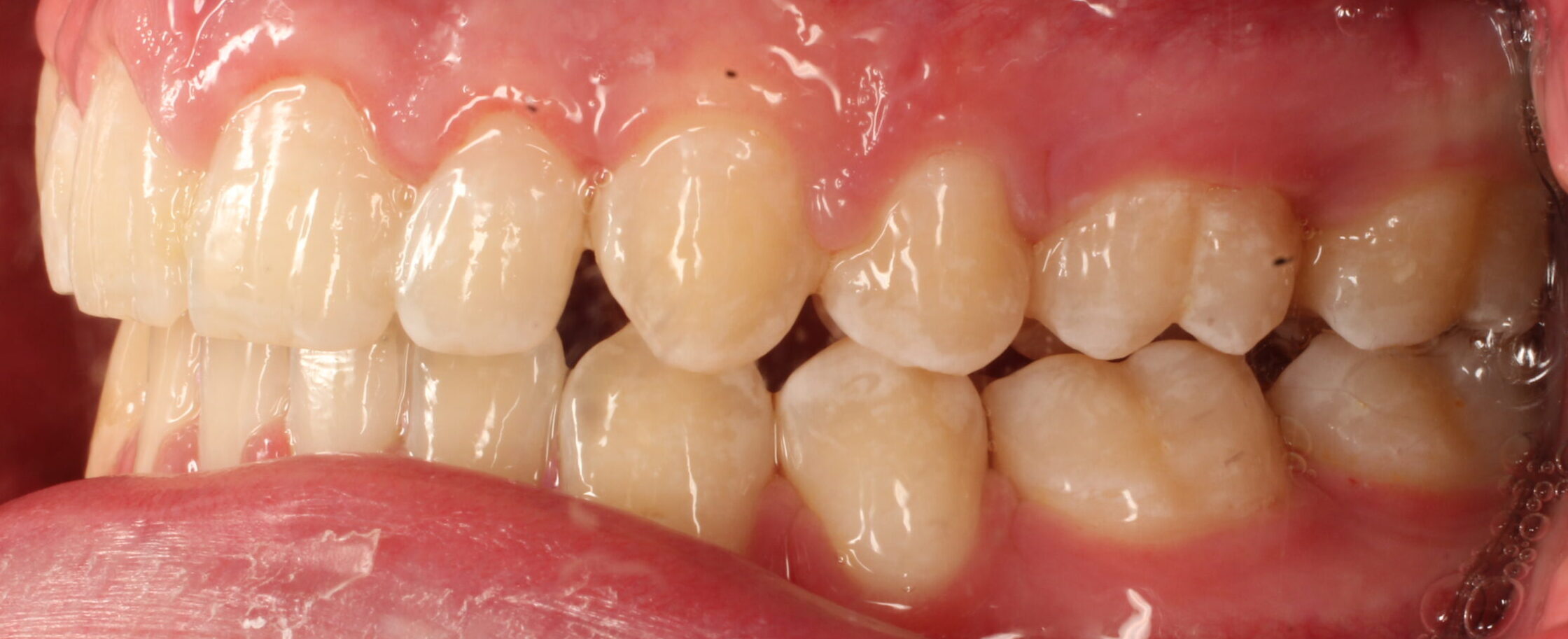
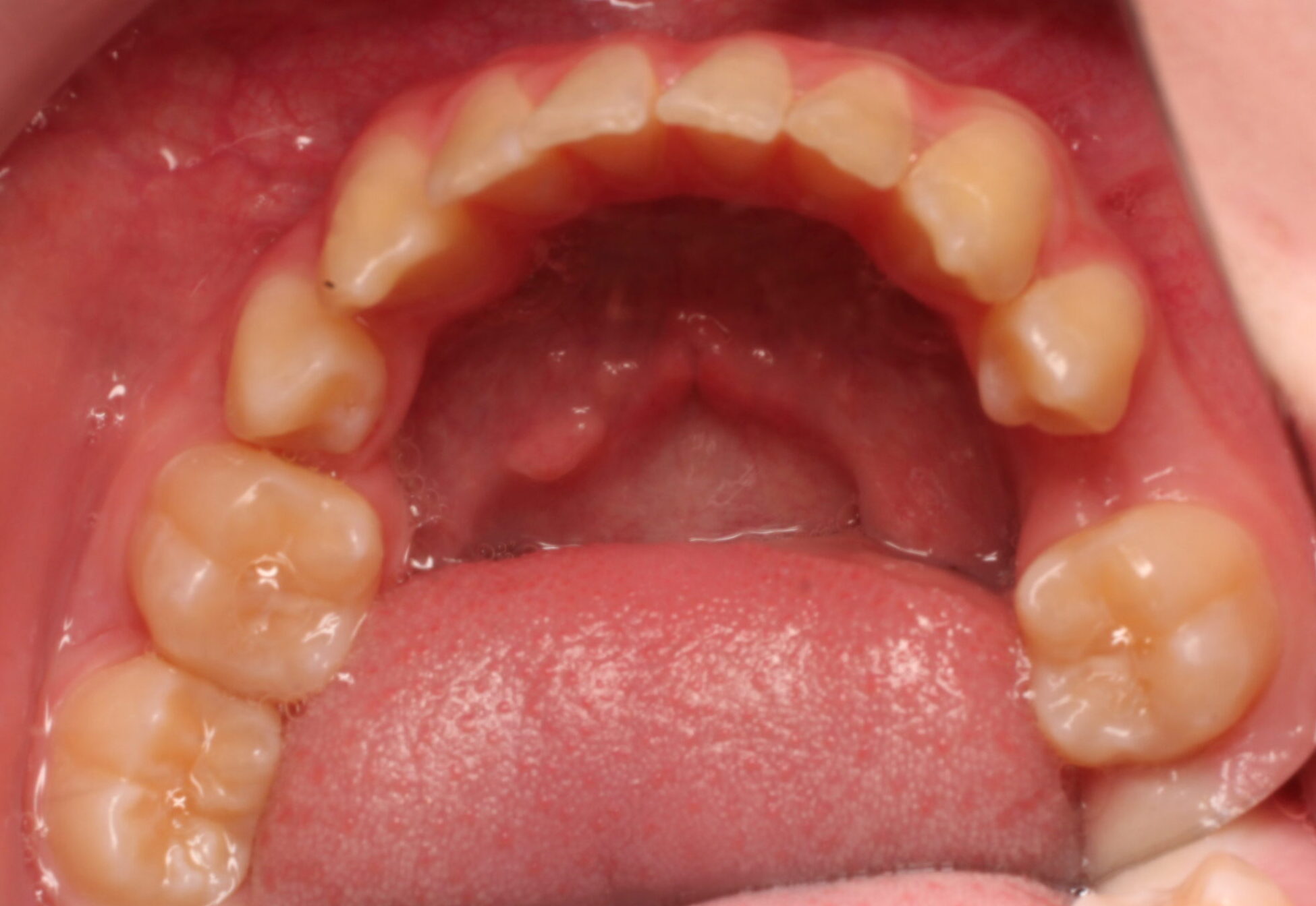
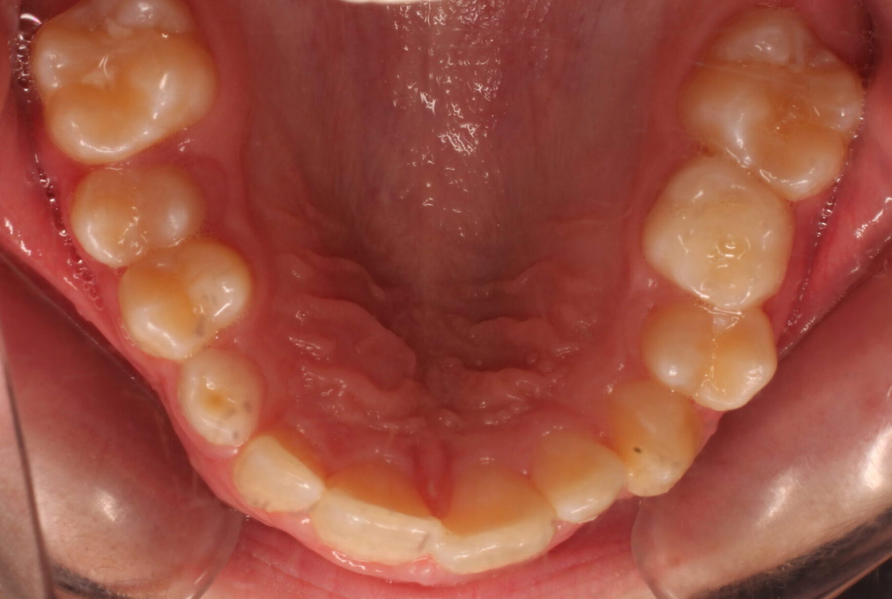
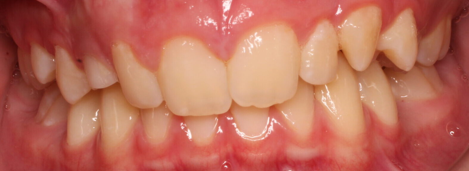
- The Trapped Adult Tooth: His upper right canine (tooth 13) was severely impacted, trapped deep within the bone, and positioned awkwardly over his front tooth.
- Arrested Root Development: High-resolution dental scans revealed a rare anomaly in the lower left quadrant—the adult second premolar (tooth 35) had completely stopped growing beneath the gums, leaving it trapped in a hazardous position extremely close to the inferior dental nerve.
- Structural Discrepancies: Matt also presented with a severe deep bite, localised crowding, retained baby teeth, and naturally small lateral incisors.
The Strategic Multidisciplinary Programme
To safely navigate these risks, our specialist orthodontist, Claire O’Sullivan, ordered a specialised Cone Beam CT (CBCT) 3D scan to map the exact relationship between the trapped teeth and the surrounding facial nerves. Armed with this data, Dr Claire O’Sullivan collaborated closely with oral surgeon Dr Marie O’Neil to orchestrate a highly precise, multi-stage treatment plan.
1. Phase One: Strategic Oral Surgery (2021)
First, Dr Marie O’Neil safely extracted the retained baby teeth and carefully removed the underdeveloped adult tooth (tooth 35) from the lower jaw, completely avoiding any trauma to the critical lower dental nerve. At the same time, the trapped upper canine was surgically exposed and fitted with a specialised gold chain mechanism, allowing our team to gently and gradually guide the tooth down into the arch.
2. Phase Two: Comprehensive Alignment & Space Management (2021 – 2024)
Once the trapped canine was successfully brought into alignment, fixed metal braces were bonded to both arches. To balance the space across his smile, our general dentist, Dr Stephen Taylor, performed comfortable extractions of his upper premolars.
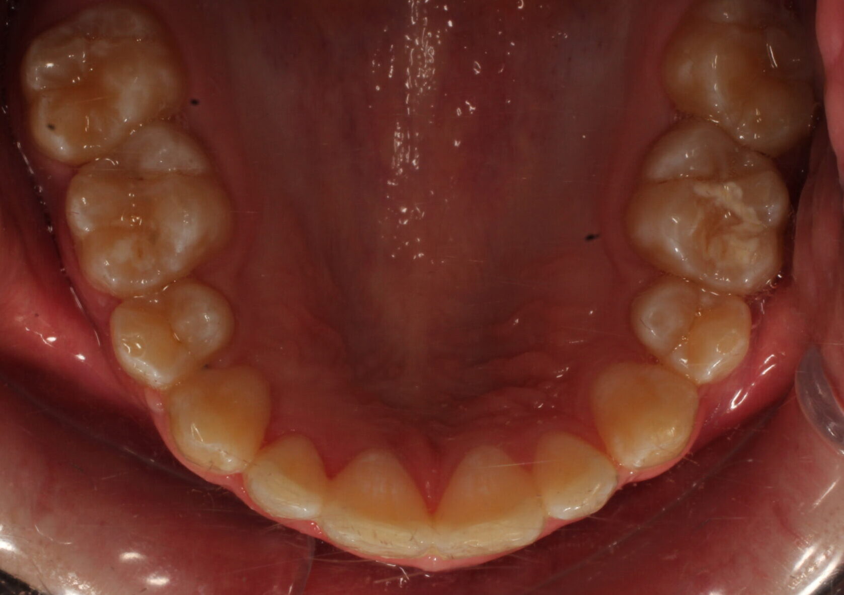
Over the next two years, our orthodontic therapists systematically closed the extraction gaps, levelled the deep bite, and coordinated his dental midlines using Class II elastics. The braces were beautifully debonded in May 2024.
3. Phase Three: Cosmetic Consultation & Active Retention
Because Matt’s upper lateral incisors were naturally small, a simple alignment would leave slight gaps next to his front teeth. Following the removal of his braces, our team initiated a comprehensive smile design consultation.
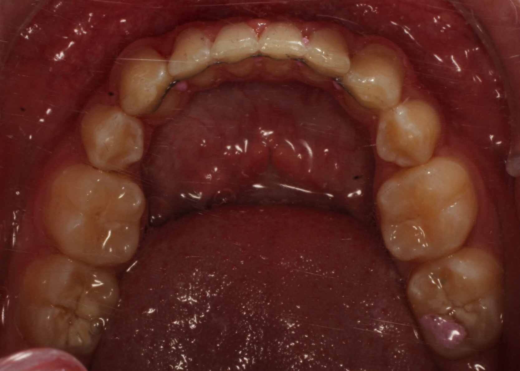
While the teeth were beautifully straight, Matt was referred to our cosmetic team for composite edge bonding to artfully build up and widen the small lateral incisors, filling the remaining spaces seamlessly. To preserve this magnificent transformation, custom-moulded upper and lower Essix retainers were delivered immediately for night-time wear.
The Phenomenal Result
Matt’s three-year journey concluded with outstanding success. By combining advanced 3D scanning with a meticulously timed orthodontic-surgical partnership, our team completely resolved multiple high-risk anomalies that could have caused long-term nerve or structural damage if left untreated. Matt finished his treatment with a structurally stable bite, excellent oral hygiene, and a balanced, radiant smile built to last a lifetime.
The Specialist Advantage: Complex cases involving impacted teeth and nerve-adjacent anomalies require a team with the advanced training to look beneath the surface. By choosing a specialist practice like Dublin Orthodontist, Matt’s family received an uncompromising standard of continuous, evidence-based care that protected his long-term health and unlocked his true smile potential.
Discover Your Child’s Customised Smile Plan
Is your child missing teeth, or has your family dentist mentioned a suspected trapped or impacted tooth? Early specialist evaluation utilising advanced diagnostics can save years of complex treatment later in life.